Interview with Alessandro Gozzi, coordinator of IIT’s ‘Functional Neuroimaging’ research line
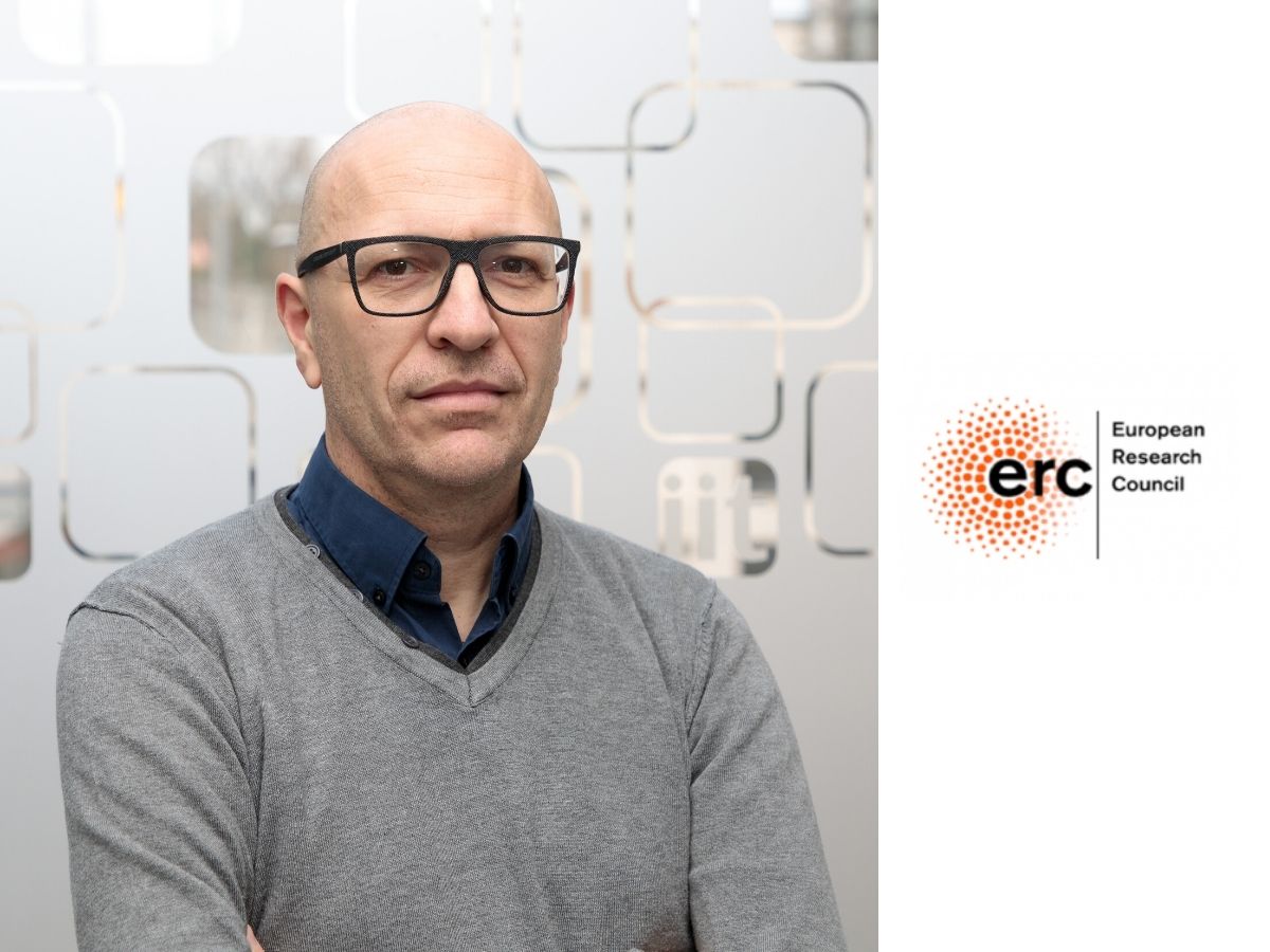
Our knowledge of the human brain is still very limited, but new information is coming to us from those who are investigating this great mystery that belongs to us. One investigator of the brain is Alessandro Gozzi, a researcher at the Italian Institute of Technology and winner of an ERC prize for the development of advanced imaging techniques to understand the functional organisation of brain activity. Thanks to his original career path, Gozzi has always wondered how research on brain diseases could concretely advance, developing innovative approaches and fully exploiting the still unexplored potential of clinical studies based on brain imaging techniques.
How far has our understanding of how the human brain works come today?
Classically, studies of brain function have supported the specialisation model: we know, for example, that there are specific areas in our brains dedicated to language, visual functions, auditory functions, memory, etc. Thus, for many decades, neuroscientists have worked around a model that describes the brain as a kind of mosaic of distinct and theoretically separable functions.
This view of the brain is the consequence of a series of investigations that are based on the measurement of a single area following a given stimulus: if neurons respond to a given visual stimulus, for example, we know that they belong to the visual area; if they do not respond, it is assumed that they do not belong there. This is the classical system.
In the last 20-30 years, however, a number of techniques have entered clinical practice to map brain activity, to photograph it and observe it as a whole. These are imaging techniques such as functional magnetic resonance imaging (fMRI) that together have revolutionised our understanding of brain activity. These studies have first of all shown that the classical model of specialisation of areas is valid and fully supported – that is, specific stimuli are able to activate specific areas. However, these studies have also shown that brain activity can be described as the intersection of two important levels of organisation: the first is that of specialisation (classical model), the second, more recent, is that of integration. The brain is therefore a very strong integrator of information. Therefore, when a specialised area is activated, it never actually activates on its own, but does so by communicating with an extensive network of many other brain areas. In other words, the brain does not function as an aggregate of mosaic tiles that can be separated from each other, but functions as a true ‘network’.
So, what do you mean when you talk about ‘functional organisation’?
Functional organisation means measuring how and how much the various areas communicate with each other, through electrical signals. It basically means being able to observe the whole brain and measure its overall network activity. Neither more nor less than we could do with other networks, for example the Internet: we can, for example, assess whether and how two or more areas (in the Internet, two computers) manage to communicate with each other and understand what kind of signals are exchanged, and how.
What do you study in your project?
We study what are referred to in the jargon as ‘connectopathies’. Apart from the theoretical aspects of how the brain works, our research touches on a very concrete point: thanks to clinical imaging studies, it has been shown not only that the brain has network activity that processes and adapts to stimuli, but, as has now been demonstrated in hundreds of studies, in all brain diseases (autism, schizophrenia, depression) this network activity is strongly altered, generating a ‘functional disconnection’. Although this is clinically evident, to date we do not know how to use this information, i.e. we do not know how to interpret it for diagnostic or treatment purposes, or to understand the causes of these diseases. What our project seeks to do, therefore, is to decode clinical imaging studies in order to understand the pathological mechanisms that lead to functional disconnection in diseases such as autism, schizophrenia etc.
What happens when there is a ‘functional disconnection’?
As in physical networks such as the Internet or the motorway network, brain networks consist of two elements: a structural substrate, namely the white matter bundles that make up the brain wiring, a kind of motorway network of the brain. The second element, which can be measured using techniques such as functional magnetic resonance imaging, is the information that these bundles transmit and thus understand how well they function (e.g. by measuring their traffic, as in road networks). When this flow of information is altered, we speak of functional disconnection, i.e. we refer to the fact that the network is not functioning as it should.
What we see in brain diseases are typically two scenarios: in a first case, which is very common in developmental diseases such as autism or schizophrenia, altered wiring is often observed. In a second case, a malfunctioning of these circuits is observed in contrast to a perfectly normal network wiring. The causes leading to this second type of malfunction still remain a mystery.
What is the aim of the project?
The ultimate goal of our project is to understand why these disconnections occur and to understand their meaning, in order to facilitate the diagnosis of these diseases, and to be able to eventually propose new forms of treatment aimed at restoring network communication between areas. In other words, our goal is to try to explain a phenomenon that we are able to see, but which we are not able to interpret to date.
How do you interpret such a phenomenon?
If we want to understand this phenomenon, we must be able to deconstruct it, manipulate it.
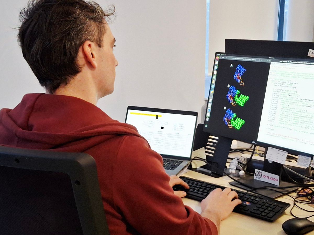
What we see is the network activity of the brain, which can be compared to an orchestra: to understand what the sound of a particular instrument is, I can listen to the piece once and then hear it again after removing the instrument, or, conversely, after turning up the volume. In this metaphor, the orchestra is the entire brain network and the instrument can be a particular area of the brain or a group of neurons, which I can silence or enhance by manipulating individual components within the system. We do both, we mute and boost the activity of specific areas, to understand their function. This type of research is done by mapping the network activity of the brain of animal models, using functional magnetic resonance imaging, the same as we use for humans. These measurements are non-invasive, as our model is simply scanned totally non-invasively for about twenty minutes, a very advanced methodology, and directly translatable to humans.
What we have seen in our studies is that a large number of genetic alterations, associated for example with autism, do not alter neuronal wiring, i.e. the anatomical structure of the white matter bundles is regular. In contrast to a preserved brain structure, which has no defects, we have, however, observed that what is greatly altered is the communication between the various areas, i.e. the way they talk to each other. There is therefore a whole series of network alterations that are not due to the fact that the network is wrong, or badly assembled, but what we suspect, and what is becoming increasingly evident, is that the network is simply dysregulated. To give an example, if I run a perfectly wired power grid at 500 volts instead of 220 volts as usual, the circuit does not work. These illnesses, therefore, do not necessarily have a problem with the assembly or wiring of the circuits, but very often they are dysregulated, they have a problem with the incorrect regulation of the signals.
Another interesting thing we do is to develop mapping to better classify developmental diseases. For example, autism is a spectrum, which is a very heterogeneous disease that does not always present itself in the same way. One of the reasons why we do not yet have any treatment is precisely because of the heterogeneity of this disease, both genetically and neurologically. In order to obtain a treatment for this type of disease, it is therefore necessary to find subcategories of patients that are more homogeneous. It has been seen in animal models that these network mappings very well define subcategories linked to particular genetic mutations associated with autism.
What is meant by subcategories?
Let us imagine patients who have a homogeneous biological or neurological dysfunction, that is, whose problems have common elements. If we think of tumours, we have realised in recent decades that there are many different types, so it is a very heterogeneous disease, for which treatments are sought according to subcategories.
In the case of autism, in many ways we are still where we were forty years ago with cancer. But we are at the beginning of a revolution, because we have now realised that there are different types of autism and imaging studies can help advance this field.
What is the challenge?
The challenge now is to understand what ‘different types’ means. There are several criteria used in this type of study: one is genetics, but it is not the only one; we are proposing another one, which does not ask what the underlying genetic mechanism is, but rather the brain dysfunction. We have seen that there are dysfunctions that cause excessive network activity, and others for which the network functions poorly. And it is unthinkable to treat two such different problems with the same drug. That is why it will be crucial to learn how to differentiate these conditions on a clinical level.
Alessandro Gozzi’s is not only a largely multidisciplinary research, in which the work carried out in the laboratory has a very high degree of technical complexity, but it is also the first study to map brain networks in rodents, and one of the few in the world that has developed methods to map brain activity between species three-dimensionally. This is why neuroscience today has started to talk about networks.


