IIT – CNR: a new form of non-invasive technology, important developments in diagnostics
(photo credits, Lorenzo Pattelli, INRiM – LENS)
Tests have been carried out on a new non-invasive light-based technique that makes innovative use of DNA nanoprobes to gain information from inside complex systems such as organs and tissue within the human body without the need for surgery or other delicate procedures.
The technique paves the way for important developments in diagnostics, as it can be applied to all cases in which the target to be reached is at a depth that renders other systems, such as X-ray or magnetic resonance imaging, ineffective due to the low spatial resolution.
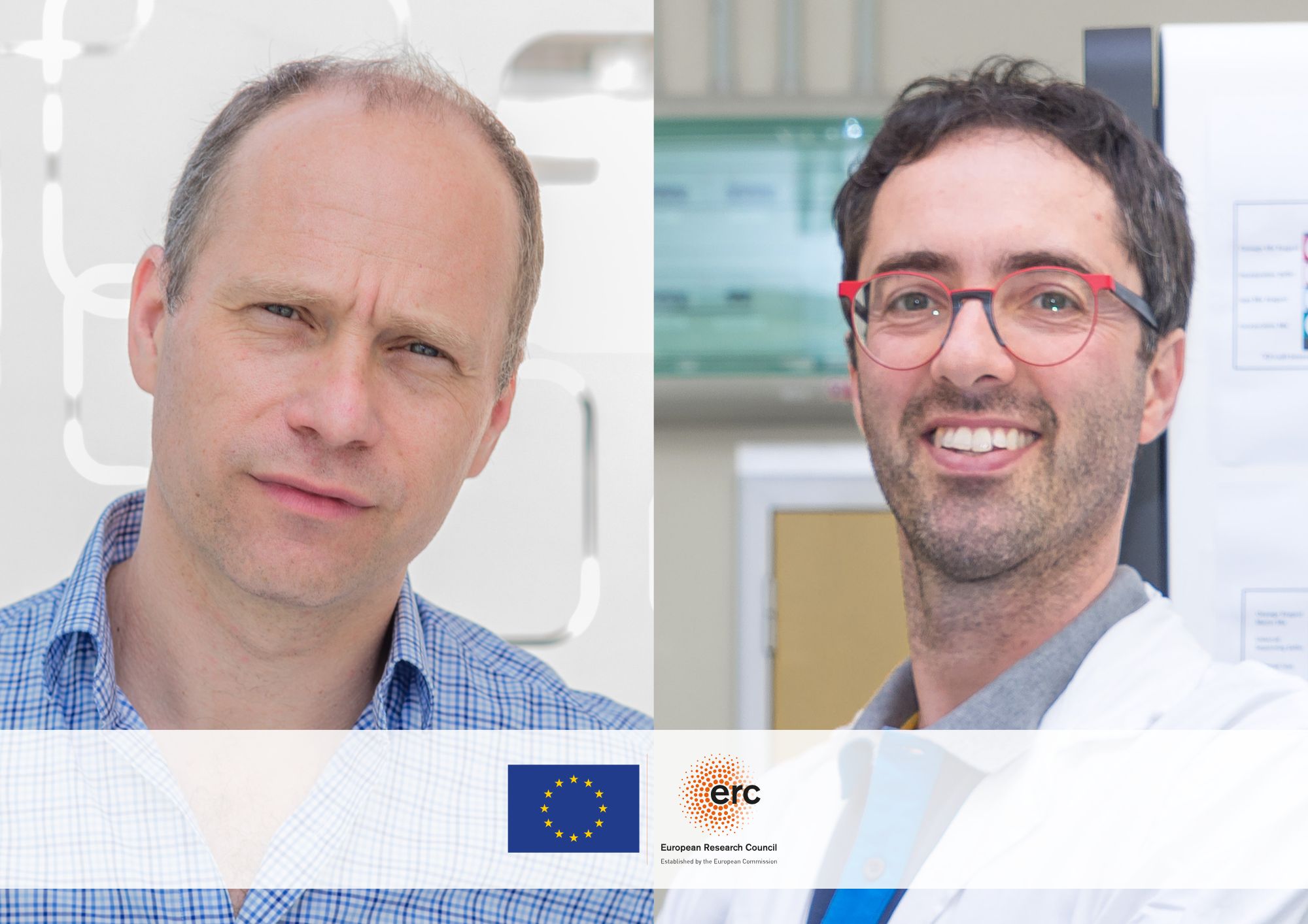
The study, which was published in the magazine Nature Communication, was conducted by Giancarlo Ruocco, coordinator of the Center for Life Nano- & Neuro-Science – CLN²S at the Italian Institute of Technology (IIT) in Rome, together with Marco Leonetti, a researcher from the Institute of Nanotechnology run by the National Research Council (CNR Nanotec) in Rome and affiliated with IIT, and with the collaboration of colleagues from the National Metrology Institute (INRiM) and the European Laboratory for Non-linear Spectroscopy (LENS).
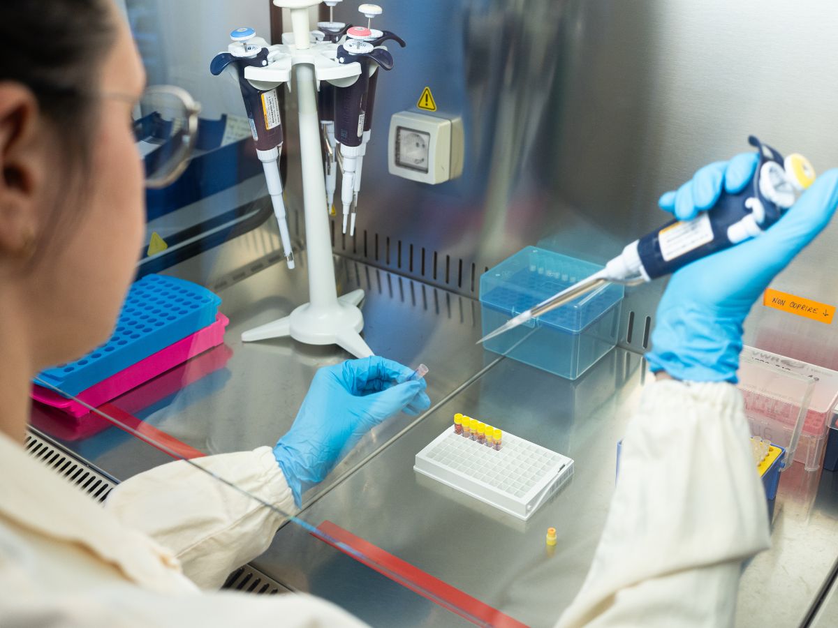
Researchers from the IIT and CNR created laboratory simulations of the diffusion of light in a complex system such as the human body, using zinc oxide, a highly reflective and biocompatible substance. The tailor-made system involves the insertion of a nanoprobe, with a diameter 10,000 times smaller than that of a human hair and capable of measuring local distortion of light. The system is illuminated with green light, which the nanoprobe detects, in turn emitting fluorescence (red light) but not providing a clear image due to the opacity of the complex system. However, by reading temporal and spatial fluctuations of the fluorescence in response to the surrounding environment, the researchers are able to recognise the presence of particular aggregates of proteins surrounding the nanoprobe, which are denser and therefore have a refraction index that differs from that of the surrounding tissue.
The results of the study represent the first steps in developing future systems for the early diagnosis of macroscopic changes in tissue that are typical of tumours or a number of neurodegenerative conditions connected to the accumulation of protein aggregates, such as Alzheimer’s.
“The nanoprobe is an object that is commonly used in microscopy and brings together the latest techniques in genetic engineering and the control of light sources”, explained Marco Leonetti, the principal author of the paper and a researcher at CNR-Nanotec, the IIT affiliate. “It behaves like a satellite sent into space, collecting information from its surrounding environment and sending it back to Earth. The nanoprobe measures the properties of light in the surrounding area and is capable of sending the results to the researchers’ measuring instruments from outside the ‘turbid barrier’. The upshot of this is that information can be gained in vivo, avoiding more invasive procedures”.
“This technique allows us to see what is happening inside tissue without having true images, but by reconstructing the system on the basis of the angle of refraction of the light”, concluded Giancarlo Ruocco, coordinator of the Center for Life Nano- & Neuro-Science at the Italian Institute of Technology (IIT) in Rome. “There are a number of implications of this discovery; one could imagine a future generation of biocompatible nanoprobes capable of providing information on the development of local changes in human tissue in inaccessible areas, as is the case with a number of neurodegenerative diseases”.
For further information: “Spatial coherence of light inside three-dimensional media”