A new hybrid device, electronic and photonic, makes it possible to study the activity of brain cells in a very precise yet minimally invasive manner, thanks to the combined application of an optical probe and micro-electrodes capable of recording the brain signal.
The integrated instrument is the result of a decade-long collaboration between the Center for Biomolecular Nanotechnologies at the Italian Institute of Technology (CBN-IIT) in Lecce, Italy, the University of Salento and Harvard Medical School (HMS) in Boston, and has been validated by research coordinated by Ferruccio Pisanello, Massimo De Vittorio and Bernardo Sabatini, the results of which were recently published in Nature Materials, one of the most relevant international scientific journals in the field of materials science.
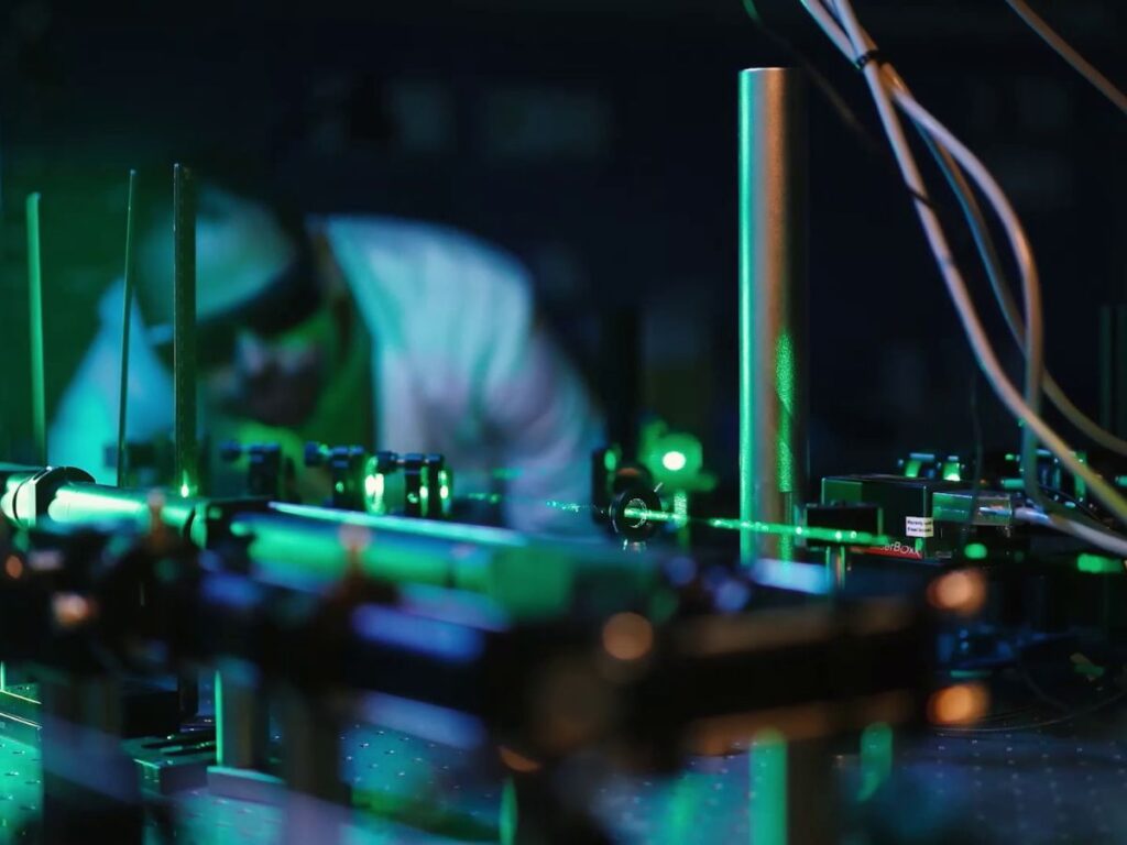
Researchers on the international team have devised a technology that allows brain activity to be read and monitored by both light and electrical coupling. This type of hybrid (electronic and photonic) device creates communication channels with the brain, allowing accurate study of the interaction between groups of neuronal cells even in areas not accessible by traditional neuroscientific investigation techniques. Specifically, the device consists of a very thin glass needle (thinner than a hair) around which a real electrical circuit has been built using an innovative micro fabrication technique. The needle has the function of transmitting optical signals to nerve cells in order to stimulate them, while the electrical circuit, in which micro-electrodes are inserted, manages to read and record the signal generated in the same cells affected by the optical beam, or in those directly connected to them.
The electrodes coupled to the probe thus make it possible to record the neuronal signal with very high spatial definition, even at the level of a single neuron. But the new instrument is also unique because, unlike other probes in which light and electrodes are coupled, in this one the light does not go to interact on the electrical circuit: that is, the phenomenon known as photo-electric noise is not encountered, and the recorded signal is therefore much “cleaner.”
The first signatories of the work, Barbara Spagnolo, Antonio Balena and Marco Pisanello of IIT and Rui Peixoto of Harvard, explain that the key feature of the device is the ability to stimulate and read nerve activity on extremely small volumes, with very little interference: “Thanks to the high degree of integration of the system, we have been able to increase the quality of the measurements to a level that was impossible to achieve until now, making the methodology finally usable for the study of the brain and its pathologies.”
In addition, Professor Massimo De Vittorio, professor of electronics at UniSalento and coordinator of the Center for Biomolecular Nanotechnologies at the Italian Institute of Technology in Lecce, and Dr. Ferruccio Pisanello point out that “The study of the interaction between cells in our brains is essential to fully understand the functioning of brain circuits, and the development of devices to stimulate and read the activity of neurons is the key that is allowing neuroscientists to analyze the mechanisms underlying the exchange of signals within the central nervous system.”
The research team is working, now, to test this technology in pre-clinical experiments, so as to understand its application in the medical field, thus enabling the development of a new approach for studying neurological and neuropsychiatric disorders.
The research was funded under projects funded by the European Union (MODEM, NanoBright, DEEPER and IN DEPTH) and the U.S. National Institutes of Health.
Link to the original paper: rdcu.be/cO4qF